
Hydrogels are three-dimensional, crosslinked polymeric networks that can retain large amounts of water, making them highly suitable for various biomedical applications. Due to their unique properties, hydrogels can mimic the natural extracellular matrix (ECM), offering an environment conducive to cell growth, proliferation, and differentiation. They are widely used in applications such as drug delivery systems, wound healing patches, and scaffolds for 3D cell cultures in tissue engineering. Additionally, their high biocompatibility and ability to adjust mechanical properties make them an ideal choice for use in implantable devices and regenerative medicine.
Gelatin-based Hydrogels
In our research, we focus on developing gelatin-based hydrogels, taking advantage of naturally derived materials like gelatin, collagen, and chitosan to improve biocompatibility and reduce the potential for foreign body reactions. By leveraging the body’s own materials, these hydrogels minimize the immune response, enhancing their effectiveness in a range of biomedical contexts. Below, we highlight our ongoing research projects involving Gelatin Methacrylate (GelMA) and GelMA-alginate interpenetrating network hydrogels.
We are exploring gelatin-based hydrogels for various biomedical applications. We are interested in hydrogels for:
- drug delivery systems
- wound healing patches
- scaffolds for 3D cell cultures and tissue engineering applications
Gelatin methacrylate (GelMA)
Gelatin Methacrylate (GelMA) is a photocrosslinkable hydrogel that offers excellent biocompatibility and the ability to tune mechanical properties for tissue engineering applications. Due to its gelatin-based structure, GelMA mimics the natural ECM, providing a favorable environment for cell culture. This versatility makes GelMA a valuable material for 3D cell cultures and regenerative medicine.

Our research focuses on optimizing the synthesis and crosslinking conditions of GelMA to improve cell viability, particularly for TC28a2 chondrocytes in a 3D culture setting. By investigating the effect of factors like GelMA concentration, photoinitiator concentration, and UV exposure time, we aim to develop a GelMA hydrogel that promotes chondrocyte survival and enhances its potential for cartilage tissue engineering.
Key findings from our study reveal that synthesis using 0.25M CB buffer results in higher methacrylation compared to PBS buffer, improving gelation efficiency. We also discovered that LAP photoinitiator outperforms Irgacure 2959, offering superior gelation properties. Additionally, we observed that increasing GelMA concentration enhances the stiffness, porosity, and swelling degree of the hydrogels. However, cell viability was negatively impacted by higher GelMA concentrations and longer UV exposure times, which must be carefully balanced for optimal outcomes.
This research provides valuable insights into GelMA optimization for biomedical applications, ensuring its potential for tissue engineering, drug delivery, and wound healing.
Read our publication related to this research:
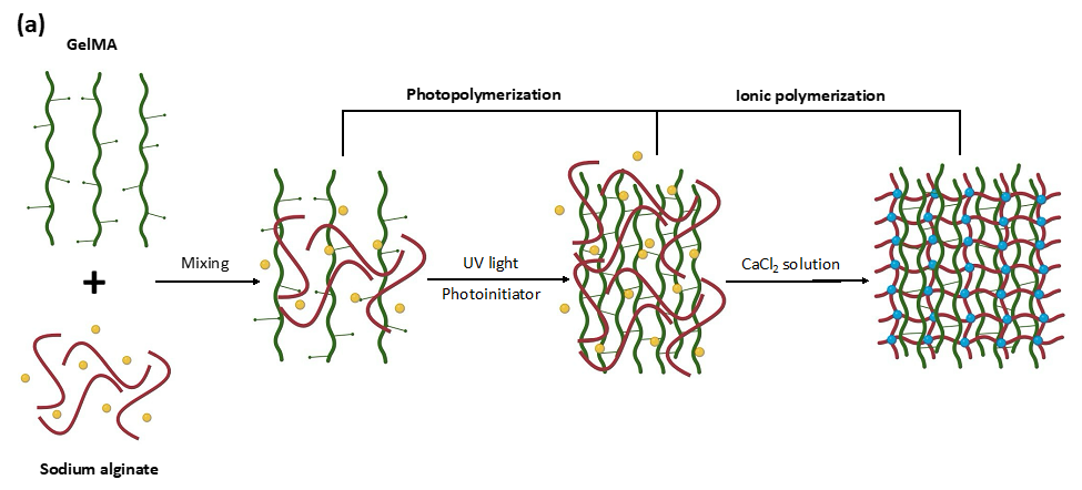
GelMA-Alginate Interpenetrating Network Hydrogel
Osteoarthritis (OA) is a chronic joint disease characterized by the degeneration of cartilage, and current treatment options do not effectively address the underlying causes. To aid in developing new therapies, we are investigating a GelMA-alginate interpenetrating network hydrogel as an in vitro model for OA. This hydrogel system combines the advantages of GelMA’s biocompatibility and mechanical tunability with sodium alginate’s water retention capacity and gel-forming abilities.
Hydrogel Composition and Properties:
Our GelMA-alginate hydrogel is used to mimic cytokine-induced features of OA, such as the breakdown of type II collagen. By incorporating MMP-13 inhibitors, we were able to demonstrate significant collagen degradation inhibition, showing the model’s potential for preclinical drug testing. Despite some inconsistencies in human cartilage explant samples, our results suggest that this hydrogel model can effectively replace human-derived tissue in the early stages of drug development.
Research Findings:
The GelMA-alginate hydrogel provides an excellent platform for drug screening and tissue engineering. The synergy between GelMA and alginate creates a biomimetic environment with customizable mechanical properties, good water retention, and an ability to support cell encapsulation and viability. This hydrogel system shows promise for cartilage tissue engineering and therapeutic agent delivery.
Read the full study here:
Q. Hu, S.L. Williams, A. Palladino, M. Ecker, Screening of MMP-13 Inhibitors Using a GelMA-Alginate Interpenetrating Network Hydrogel-Based Model Mimicking Cytokine-Induced Key Features of Osteoarthritis In Vitro. Polymers 2024, 16, 1572. https://doi.org/10.3390/polym16111572
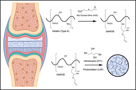
Thiol-Clickable Gelatin-Based Hydrogels for 3D Cell Cultures and Tissue Engineering
Thiol-clickable hydrogels, which leverage thiol-ene click chemistry, are gaining significant attention in the field of biomedical materials due to their customizable properties, making them ideal candidates for tissue engineering and drug delivery applications. When combining gelatin with thiol-ene chemistry, two primary approaches are used: (1) introducing thiol groups to the gelatin backbone, or (2) functionalizing the gelatin with alkene groups. Both approaches allow for the formation of hydrogels that can be crosslinked under mild conditions using thiol monomers.
In most studies, gelatin is thiolated using thiol compounds such as cysteine or norbornene, allowing the pending thiol groups to crosslink the gel. However, there are fewer examples where gelatin-based hydrogels are crosslinked using thiol monomers. In one example, gelatin (Gel) is functionalized with allyl glycidyl ether (AGE), creating allyl functional groups. These functionalized gels are then crosslinked with dithiothreitol (DTT) to create the hydrogel matrix. Although this approach offers a promising path to custom gel properties, it has mostly been limited to the use of dithiothreitol as the crosslinking thiol moiety.
Our research expands upon this approach by exploring a broader range of thiol monomers with different chain lengths, backbones, and functional groups. By incorporating tri- and tetra-functionalized thiol monomers, we can more precisely manipulate the network structure, stiffness, pore size, and nutrient transport properties of the hydrogels. This tunability opens the door to customizing hydrogels for various biomedical applications, including the development of 3D cell cultures that better mimic the extracellular matrix (ECM) of human tissues.
Specifically, we aim to develop gelatin-based hydrogels that replicate the mechanical and thermomechanical properties of cartilage tissue, which is notoriously difficult to mimic with existing hydrogel systems. Current hydrogels either lack the necessary mechanical properties to resemble cartilage or fail to support 3D cell cultures effectively. By combining natural gelatin-based hydrogels with synthetic thiol-ene-based polymers, we hypothesize that we can create a bioinspired material that supports cell growth and differentiation, while offering superior mechanical properties and nutrient transport.
To achieve this, we will utilize allylated gelatin (GelAGE) as the base material and combine it with various thiol monomers to optimize crosslinking conditions. By varying the thiol monomer choice—specifically, the chain length, number of functional groups, and chemical structure—we can achieve a matrix that mimics the natural ECM of cartilage more closely. This approach will allow us to fine-tune the pore size, hydrophilicity, and mechanical strength to match the requirements for cell viability and differentiation.
Our research will compare the mechanical properties and cell growth capabilities of these engineered hydrogels against natural cartilage to ensure the system is optimized for in vitro 3D cell cultures. We expect this thiol-clickable hydrogel system to be an invaluable tool in disease modeling, drug screening, and the development of cartilage tissue engineering applications.
These research projects are part of our broader effort to optimize and develop hydrogel-based materials that can be used for a range of biomedical applications, from tissue regeneration to drug delivery and wound healing. Our work focuses on creating customized hydrogels that enhance biocompatibility, mechanical properties, and cellular interactions, offering new possibilities for personalized medicine and regenerative therapies.